Medication Techniques
Learning Objectives
- Be able to locate and gently occlude the following veins on a dog:
- Cephalic
- Saphenous
- Jugular
- Be able to locate an appropriate site for intramuscular injection.
- Be able to locate an appropriate site for subcutaneous injections.
Live dogs will be used to locate anatomic sites for injections and venipuncture in small animal patients. NOTE – the injections or venipuncture will not be performed in this laboratory on dogs, but the topographical anatomy relevant to medication techniques will be taught.
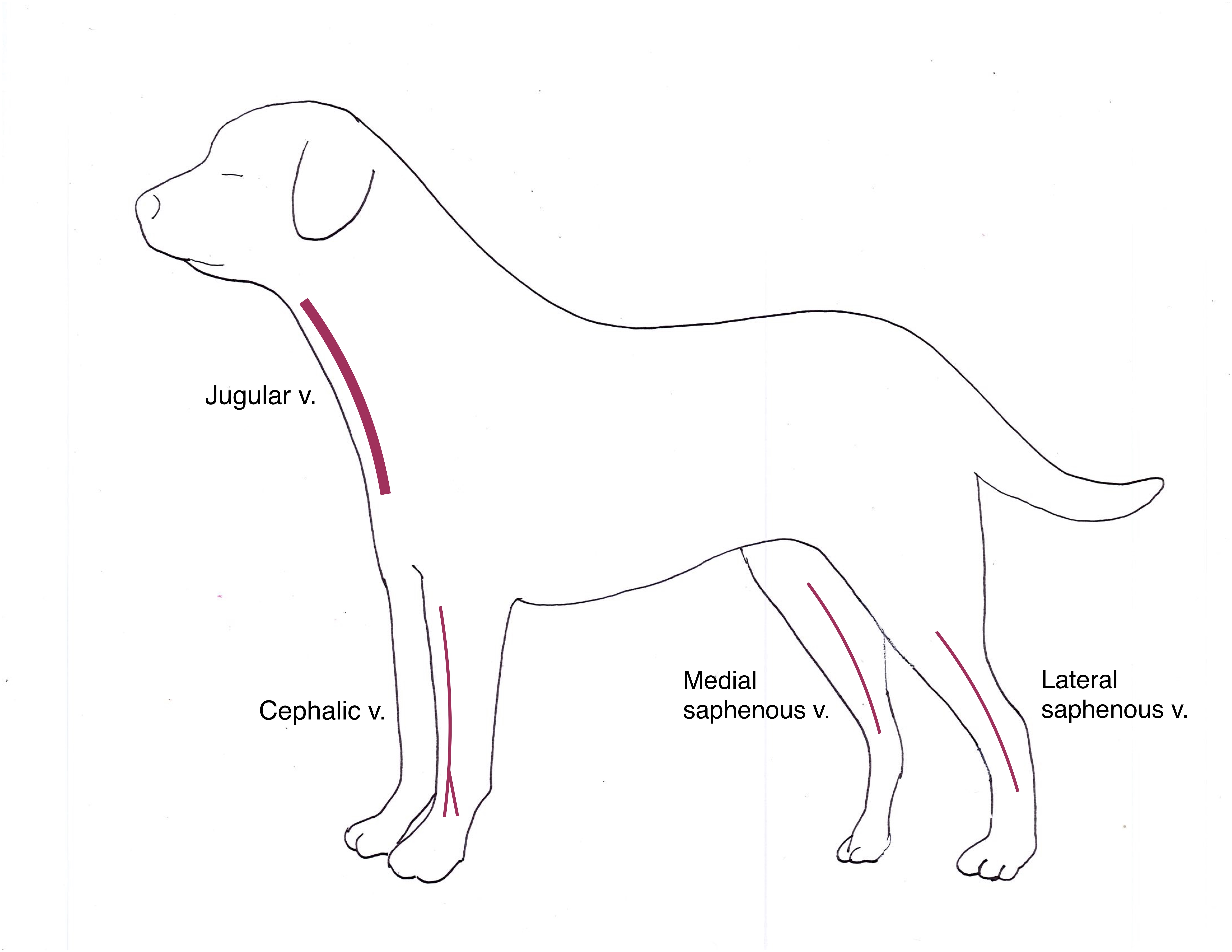
Peripheral veins
Using the diagram and the in-lab demonstration as a guide, locate the approximate location of the following veins:
- Cephalic vein (forelimb)
- Lateral saphenous vein (hind limb) – typically used only in dogs
- Medial saphenous vein (or “femoral” vein; hind limb) – typically used only in cats
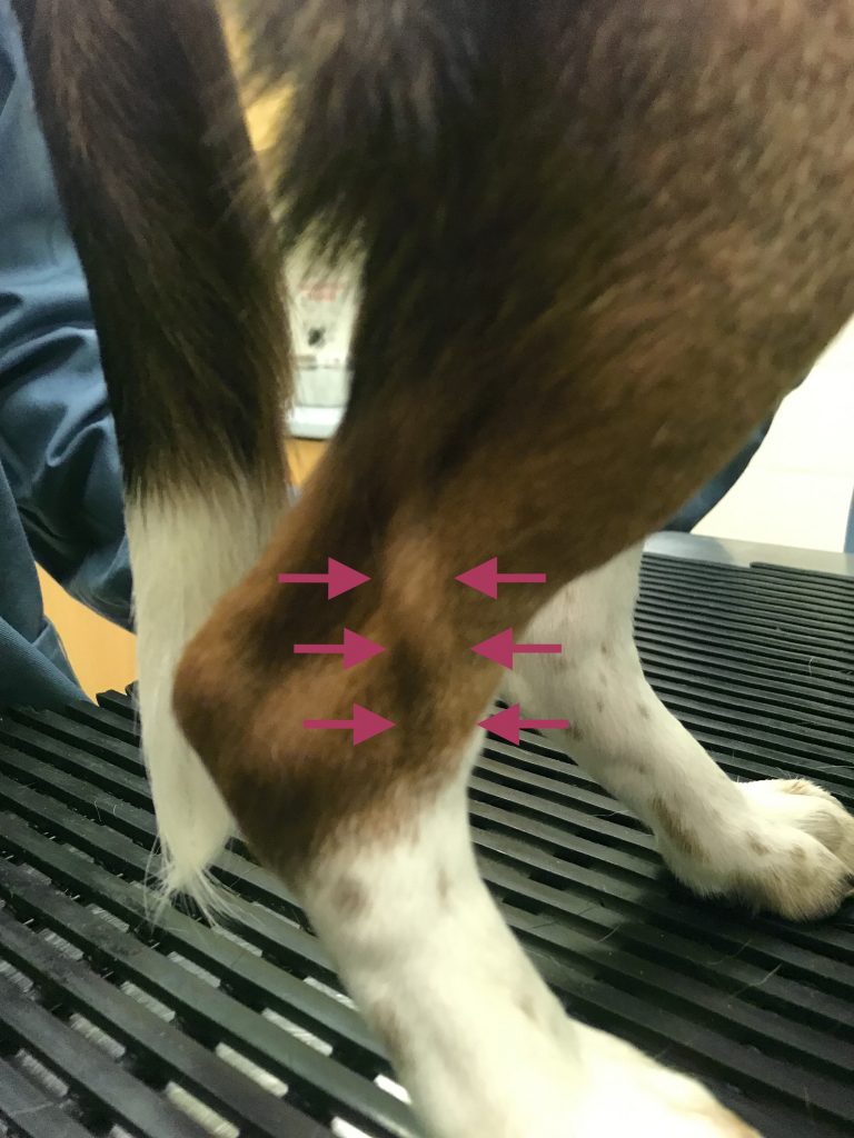
Practice holding off the limb proximal (above) the saphenous or cephalic vein. Form a circle with your hand in this location to occlude the vein distal (below) where you are holding off. You should see the vein raise up slightly when it is being occluded or held off. Palpate the vein once it is occluded to estimate its location and diameter.Veins are occluded for a brief period to allow for blood sampling or catheter placement.
Placing a small amount of rubbing alcohol over the vein will help with visualization. Alternatively, a small patch of fur can be shaved in the lab to facilitate visualization.
Jugular vein
The jugular vein is a large vein used to collect blood. It can be more difficult to visualize than a peripheral vein as it is not as superficial. Sitting restraint is often used to facilitate jugular venipuncture.
The jugular veins run parallel and lateral to the trachea. Occlude the jugular vein by placing your thumb horizontally into the jugular furrow at the level of the thoracic inlet.
The vein can be palpated once it is occluded. To determine if you are palpating the jugular vein, remove pressure from the jugular furrow – the vein should no longer be palpable. Occlude the jugular furrow again, and the vein should become palpable.
Intramuscular (IM) Injection Site
Intramuscular injections are used to administer small volumes of medications. Several sites can be used to inject into the muscle. In this lab, we will examine the epaxial muscles as an IM injection site. Epaxial (lumbodorsal) muscles are on either side of the lumbar vertebrae. To identify the epaxial muscle site for IM injection, first palpate the dorsal spinous process (midline). Next, palpate the muscles along the lateral aspect of the lumbar spine to identify the injection site.
Subcutaneous (SQ, or SC) Injection Sites
Subcutaneous injections are performed to administer some medications (if labelled for SC use), most vaccinations (check label), and can also be used to administer fluids.
SC injections are often administered over the dorsal region of the neck or back, where a “skin tent” can be gently and easily grasped. (One exception to these sites is for feline vaccination – recommended protocols are to inject SC over the distal aspects of the limbs). To locate a SC injection area, gently grasp a skin tent using your thumb and middle finger. Using your index finger of the same hand, palpate the area of the tented subcutaneous space to confirm injection site.
