Spinal Reflexes
The spinal reflexes evaluate the integrity of the sensory (afferent) and motor (efferent) component of the reflex arc and the influence of descending motor pathways. They are simple and non-conscious reactions (does not reach cerebral cortex) trigger by a stimulus. They allow us to assess the status of specific peripheral nerves and spinal cord segments involved in their reflex arc. We assess:
- PATELLAR REFLEXES: in both pelvic limbs
- FLEXOR (WITHDRAWAL) REFLEXES: in all four limbs
- PERINEAL REFLEX
- CUTANEOUS TRUNCI REFLEX
Patellar Reflexes:
Position the patient in lateral recumbency while placing one hand under the thigh or the foot to support the limb. With the other hand, briskly hit the patellar tendon with a reflex hammer. Normal response is a single, fast extension of the knee. This reflex assesses the integrity of:
- Femoral nerve(sensory/afferent and motor/efferent)
- L4-L6 spinal cord segments
Flexor Reflexes:
With the limb extended, the interdigital skin or the distal phalanx is pinched with our fingers. Normal response is flexion of the hip, knee, and hock in the pelvic limbs, and the shoulder, elbow, and carpus in the thoracic limbs. This reflex assesses the integrity of:
In the pelvic limbs:
-
- Sciatic nerve (sensory and motor) when stimulating lateral toe, or femoral nerve (sensory) and sciatic nerve (motor) when stimulating medial toe.
- L6-S2 spinal cord segments
In the thoracic limbs:
-
- Brachial plexus
- C6-T2 spinal cord segments
Perineal Reflex:
Lightly touching the skin just laterally to the perineum or the actual perineum in both sides. Normal response is tail twitching/flexion and contraction of the anal sphincter. It assesses:
- Pudendal nerve(sensory and motor)
- S1-S3 spinal cord segments
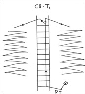
Cutaneous Truci Reflex:
The skin just lateral to the spine is lightly pinched in both sides starting just cranial to the wings of the ileum around L5. Normal response is a twitch of the skin over the thorax and abdomen in both sides, due to the contraction of the cutaneous trunci muscle, independently of the side pinched. If no response is elicited, advance to pinch more cranially one vertebral level at a time until a response is obtained. From the dermatome pinched, the sensory nerve from the skin enters the spinal cord 1-2 vertebrae cranial to the level tested and then ascends cranially and synapses bilaterally at C8-T1 spinal cord segment where the lateral thoracic nerve exits the spinal cord through the brachial plexus and innervates the cutaneous trunci muscle. An obvious cutoff point suggests spinal cord lesion one or two vertebrae cranially.