Head and Neck
Skull
While the examiner is holding the head, the shape and symmetry of the skull can be assessed for shape and symmetry. The skull can also be checked for open fontanelles in certain breeds such as Chihuahuas. An open fontanelle is an opening in the skull on the top of the head where the parietal and frontal bones come together. This defect can be (but not always) associated with hydrocephalus.
Eyes
Prior to the eye examination, appreciate facial symmetry. Look for asymmetry between the right and left side of the face and paying attention to the region of the eyes:
- Are both eyes open to the same degree (winking or squinting noted)?
- Is the third eyelid more noticeable in one eye versus the other (pink membrane in medial cornea of the eye)?
- Is the position of each eye normal within the orbit (sunken, forward, off side)?
Discharge from the eyes and nose should be noted and described, if present.
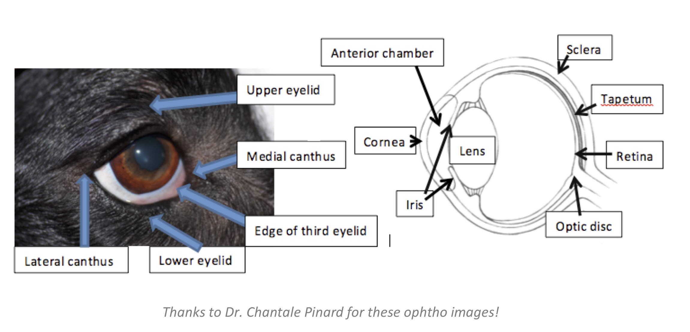
Each eye is examined separately from the front to the back with a bright light source. Be aware of the light source and for the sake of the dog’s comfort, do not shine into the eye for a prolonged period. Note the following structures on your examination:
- Upper and lower eyelids: skin covered
- Third eyelid: pink mucous membrane structure coming from medial canthus
- Conjunctiva: usually transparent with few small blood vessels
- Sclera: white part of the eye beneath conjunctiva
- Cornea: should be transparent
- Anterior chamber: empty space between cornea and iris
- Iris: pupil in dog should be round; thin oval in cats.
- Lens: should be transparent and let the tapetal reflection through when shining a light
- Tapetal reflection: “eye shine” that dogs and cats have (humans have a “red reflection”)
- Further ophthalmologic examination techniques will be taught in Clinical medicine III
Ears
The external pinnae should move in response to sound and a light touch on the interior side. Gently raise the external pinna to examine the exterior portion of the ear canals; an otoscope is used to examine the internal canal and tympanic membrane, if indicated, and this will be taught in a dermatology laboratory later in the curriculum.
Nose
The bridge of the external nose is examined for shape and symmetry. The planum nasale is examined for color and erosions. The nares are examined for discharge and patency. If there is a concern regarding nasal disease or nasal patency, a microscope slide or small piece of cotton fluff can be placed in front of each nare. If the name is patent, condensation will appear on the slide or the cotton fluff will move with air passage. The interior of the nasal cavity cannot be examined without heavy sedation or general anesthesia.
Mouth
The mouth should not be examined in an animal with neurological signs and an unknown vaccination history (primarily rabies vaccination).
Open the mouth by grasping the maxilla with one hand and the mandible with the other. Resistance to opening the mouth is often behavioural, but could indicate pain in or around the mouth.
Examine the appearance of following structures to gain an appreciation for normal appearance:
- The lips and mucocutaneous junctions.
- Mucous membranes (colour, feel).
- Gums, hard palate, soft palate (e.g., rule out cleft palate in neonates).
- Teeth.
- Tongue (appearance, movement).
Mucous membranes can be discoloured due to disease (cyanosis, icterus, hyperemia) and if the animal is dehydrated the membranes will be dry to the touch. Excess panting can also cause dry membranes; this is not the only marker of dehydration.
Capillary refill time can be performed by gently pressing your fingertip on a pink or non-pigmented area of the gums to blanch the capillaries and then observing for refill time. Normal is <2 seconds. A slow CRT (> 2 seconds) may be indicative of poor peripheral perfusion, however, it is not a very sensitive test.
The teeth are visualized and can be palpated for pain or looseness. The cheeks are retracted from the teeth, and the buccal surface of the teeth and the cheek pouch are examined.
Use a cotton swab or tongue depressor to examine under the tongue for masses, foreign bodies, or other abnormalities.
The tonsils are not normally visualized, but may be evident if they are inflamed. They can be visualized by depressing the base of the tongue.
The first video shows examination of the closed mouth; the second video shows examination once mouth is opened:
Salivary glands
Parotid glands are located at the junction of the head and neck at the base of the ear and may not always be palpable. The mandibular gland lies caudal to the angle of the jaw, and should always be palpable. The sublingual gland is closely related to the anterior end of the mandibular gland and may not always be palpable.
In this laboratory, pay particular attention to differentiating mandibular salivary glands from lymph nodes – details below in lymph node section. The video below shows palpation technique for the mandibular salivary glands.
Neck
Palpate along the dorsal, lateral, and ventral surfaces of the cervical region for any pain, asymmetry or other palpable abnormalities. Flex and extend the neck gently to determine if there are any areas of pain. Further movement of the neck will be discussed in the Detailed Neurological Examination.
Palpable structures in the ventral cervical region include:
- Larynx
- Trachea
- Jugular vein (when vein is held off at the thoracic inlet)
Coughing on palpation of the trachea can indicate tracheitis.
The thyroid gland is usually not palpable unless enlarged. (e.g., thyroid mass, feline hyperthyroidism).
The esophagus is not palpable unless there is a large mass or foreign body within it.
