Milk Production
6 Milk Biosynthesis
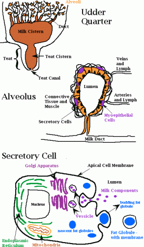
Milk is synthesized in the mammary gland. Within the mammary gland is the milk producing unit, the alveolus. It contains a single layer of epithelial secretory cells surrounding a central storage area called the lumen, which is connected to a duct system. The secretory cells are, in turn, surrounded by a layer of myoepithelial cells and blood capillaries.

The raw materials for milk production are transported via the bloodstream to the secretory cells. It takes 400-800 L of blood to deliver components for 1 L of milk.
- Proteins: building blocks are amino acids in the blood. Casein micelles, or small aggregates thereof, may begin aggregation in Golgi vesicles within the secretory cell.
- Lipids:
- C4-C14 fatty acids are synthesized in the cells
- C16 and greater fatty acids come directly from feed or are preformed as a result of rumen hydrogenation and are transported directly from the blood to the secretory cell
- Lactose: milk is in osmotic equilibrium with the blood and is controlled by lactose, K, Na, Cl; lactose synthesis regulates the volume of milk secreted
The milk components are synthesized within the cells, mainly by the endoplasmic reticulum (ER) and its attached ribosomes. The energy for the ER is supplied by the mitochondria. The components are then passed along to the Golgi apparatus, which is responsible for their eventual movement out of the cell in the form of vesicles. Both vesicles containing aqueous non-fat components, as well as lipid droplets (synthesized by the ER) must pass through the cytoplasm and the apical plasma membrane to be deposited in the lumen. The milk fat globule membrane is comprised of the apical plasma membrane of the secretory cell, and the plasma membrane is continually renewed by the vesicles.
Milking stimuli, such as a sucking calf, a warm wash cloth, the regime of parlour etc., causes the release of a hormone called oxytocin. Oxytocin is released from the pituitary gland, below the brain, to begin the process of milk let-down. As a result of this hormone stimulation, the muscles begin to compress the alveoli, causing a pressure in the udder known as letdown reflex, and the milk components stored in the lumen are released into the duct system. The milk is forced down into the teat cistern from which it is milked. The let-down reflex fades as the oxytocin is degraded, within 4-7 minutes. It is very difficult to milk after this time.

