Kidney Basic Function and Anatomy
The anatomy of the kidney can be used to understand how it filters waste products from the body and why hormones can impact different areas throughout the kidney. They can be found at the back of your abdomen, on either side of your spine and are protected by the rib cage. The functional unit of the kidney is known as the nephron and its vasculature.
Learning Outcomes
In this section you will learn…
- Describe and label the anatomical structures of the kidney and understand their relationship to the functions of the kidney.
- Identify the structures and divisions of the nephron.
- Understand the general anatomy of the macula densa.
Anatomy of the Kidney
When studying the anatomy of the kidney it is helpful to keep in mind the functions of the kidney:
- Clear the blood plasma of unwanted substances – there is filtration of substances from the blood into the kidney, then reabsorption of important substances back into the blood, unwanted substances remain in the kidney to eventually be eliminated from the body
- Volume regulation – through renal blood flow (RBF)
The diagram below is a cross-sectional view of the kidney. Do not spend too much time trying to memorize these structures. This image is best used to understand where different processes and functions are occurring.
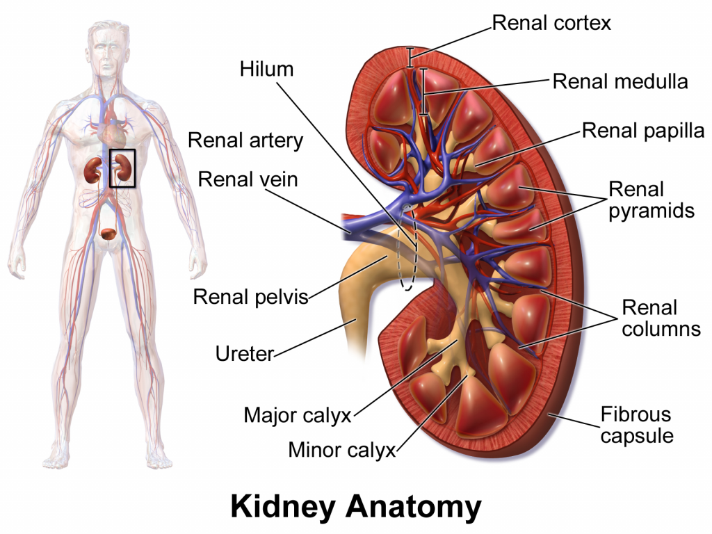
The kidney is composed of various internal structures. The gross anatomy of the kidney can be divided into an outer region called the renal cortex, and an inner region called the renal medulla which consists of renal pyramids and the renal columns. The renal pyramids are triangular in shape and renal columns are found organized within these pyramids. Together, the renal pyramids and renal columns comprise the lobes of the kidney.
The minor calyces extend from the renal pyramids and converge to form the major calyces. The major calyces then drain into the renal pelvis, which collects the urine produced by the kidney. The urine then exits the renal pelvis through the ureters and is then stored in the bladder until excretion.
The renal capsule is a fibrous layer, surrounding the entire outer surface of the kidney, which helps to cushion and protect the kidney. The renal capsule is also covered with a shock-absorbing layer of adipose tissue and a tough layer of fascia that further helps to protect the kidney and anchor it to the posterior abdominal wall. [1]
Tips from Past Students
When reading through this unit keep trying to make connections between the various sections.
Anatomy of the Nephron
The nephron is the functional unit of the kidney. It is functionally compartmentalized, meaning it is a long tube with different parts that each have other functions.
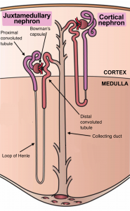
The diagram below shows the two different types of nephrons in the kidney: a juxtamedullary nephron and a cortical nephron. Notice how the juxtamedullary nephron has a Loop of Henle that extends deep into the medulla, whereas the cortical nephron is primarily in the cortex. We mainly focus on juxtamedullary nephrons for the remainder of this unit. [2]
The nephron is the functional unit of the kidney. Nephrons are located in the renal cortex and the renal medulla of the kidney and there are many nephrons located throughout the cortex and the medulla. Urine production begins in the nephron and then it drains into the collecting ducts (renal papillae are bundles of collecting ducts). The urine is then passed into the minor calyces and then the major calyces, where it drains into the renal pelvis and then the ureter for excretion.
Nephrons accomplish 3 principle functions- filtration, reabsorption, and secretion. The tubular network discerns nutrients from wastes, and selectively replenishes the blood with water, salt, and nutrients that have entered the filtrate. Additionally, nephrons have some secondary functions, which include blood pressure regulation ( via renin production-see below), red blood cell production and calcium absorption.
There are two different types of nephrons, cortical nephrons that do not extend past the renal cortex, and juxtamedullary nephrons that have a loop of henle that extends deep into the renal medulla. The capillary network that surrounds the loop of Henle in the juxtamedullary nephrons, is called the vasa recta.
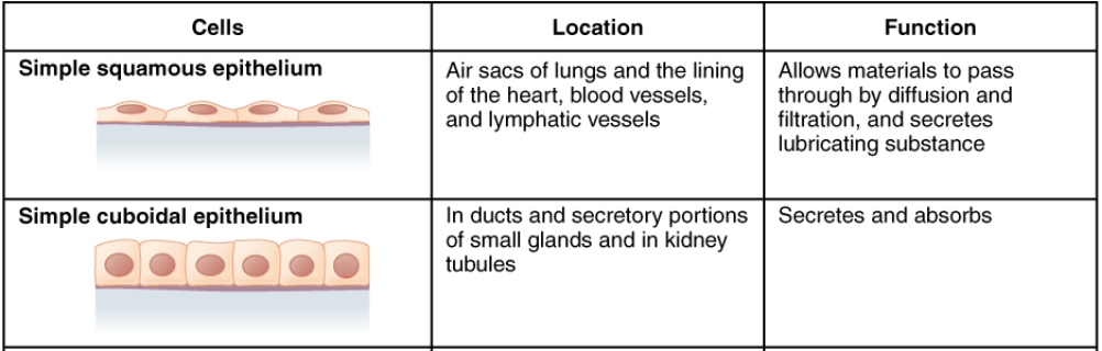
Different types of epithelium exist throughout the nephron. The epithelium of the cell influences the function of the tubule. The following chart helps summarize the function of Simple squamous epithelium, and simple cuboidal epithelium. [3]
The chart below outlines the different types of epithelium cells that can be found in the nephron. It is important to understand the differences between the different cell layers but no need to memorize them. This learning object is beyond the course level. [4]
Classification of epithelium in the nephron:
Renal Corpuscle
The renal corpuscle can be found in figure 4 and consists of the glomerulus (a high-pressure capillary bed) surrounded by the Bowman's capsule (the first section of the nephron tubule). Afferent arterioles lead into the glomerulus, and efferent arterioles lead away from the glomerulus which can be seen in figure 4. The Bowman’s capsule captures and directs filtrate from the glomerular capillary bed to the proximal convoluted tubule (PCT) of the nephron. [5]
Did you know the capillaries are fenestrated? This maximizes the amount of liquid that can leave the blood to become filtrate.
The figure below outlines the path the filtrate follows through the nephron to be excreted in the urine. Along this path, many different particles are absorbed or lost which depends on the location of the filtrate in the nephron.

Proximal Convoluted Tubule (PCT)
As seen in figure 4, the PCT filtrates the contents from the Bowman’s capsule which is a diluted solution of water, salts, nutrients, and wastes. Most of these substances are essential nutrients that need to be returned to the blood. It is called a ‘convoluted’ tubule due to its twisted structure. Simple cuboidal cells form this tubule with prominent microvilli on the luminal surface, forming a brush border. The microvilli create a large surface area, maximizing the absorption and secretion of solutes. The majority of nutrients found in the filtrate are reabsorbed into the blood via the PCT through active transport. [6]
Loop of Henle
The loop of Henle is a hairpin-like structure, with ascending and descending portions, which consists of both thick and thin portions. The loop of Henle can be divided into three different functional sections: the thin descending limb and the thick and thin ascending limbs, which can also be found in figure 4.
The thick descending limb of the loop of Henle contains simple cuboidal epithelium similar to the PCT and is responsible for actively transporting salts back into the blood. The thin portions of the descending and ascending limbs consist of simple squamous epithelium and are responsible for the diffusion of water back into the blood. The thick ascending limb contains simple cuboidal epithelium, similar to the distal convoluted tubule (DCT) (see below for more information).
The space within the kidney that the loop of Henle occupies classifies whether the nephron is a cortical nephron or a juxtamedullary nephron. Cortical nephrons have a short loop of Henle that does not extend far into the medulla. In contrast, the loop of Henle of juxtamedullary nephrons extends deep into the medulla. [7]
Distal Convoluted Tubule (DCT)
The distal convoluted tubule in figure 4, is a twisted structure similar to the PCT that consists of cuboidal epithelium. However, the DCT is shorter in length than the PCT and has fewer microvilli on the apical surface. There are several mitochondria in the cuboidal cells of the DCT, so they can pump ions against their concentration gradients (this requires a lot of energy). The DCT responds to hormonal signals that regulate urine composition. [8]
Collecting Ducts (CD)
The collecting duct is continuous with the nephron, but it is not considered to be a part of the nephron, also in figure 4. Each collecting duct has various nephrons that are merging with it. Distally, the collecting ducts merge to form the terminal ducts and ureters that drain into the bladder. The collecting ducts are lined with simple squamous epithelium that contain receptors for Antidiuretic Hormone(ADH). ADH increases the concentration of aquaporins in the CD, increasing water reabsorption. [9]
Ever wonder how many aquaporins are in the human body? There are at least 10 types and 6 of these aquaporins are found within the kidney.
The table below is a great tool to use once you have understood what each part of the nephron is capable of doing. Don’t forget that the late distal convoluted tubule, cortical and medullary collecting duct have an increase in water permeability with the presence of ADH-V, which you will learn more about in the hormonal subunit!
Table 1: Summary of Tubule Characteristics in the Kidney
| Location in the Nephron | Water Permeability | NaCl Permeability | Urea Permeability | Active NaCl Transport |
|---|---|---|---|---|
| Thin descending loop of Henle | high | low | low | 0 |
| Thin ascending loop of Henle | 0 | high | low | 0 |
| Thick ascending loop of Henle | 0 | low | 0 | high |
| Early Distal Convoluted tubule | 0 | low | 0 | low |
| Late Distal Convoluted tubule | 0 | low | 0 | low |
| Cortical Collecting duct | 0 | low | 0 | low |
| Medullary Collecting duct | low | low | moderate | low |
Macula Densa
The figure below shows a cross-section of the macula densa. As you can see it is tightly packed with specialized cells and while you are not expected to reproduce this, you do need to know where it is located and how it interacts with afferent arterioles. [10]
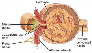
Lying just outside Bowman’s capsule and the glomerulus is the juxtaglomerular apparatus (JGA). At the juncture where the afferent and efferent arterioles enter and leave the Bowman’s capsule, the initial part of the distal convoluted tubule comes into direct contact with the arterioles. This area is known as the macula densa. This cluster of cells monitors the fluid composition that is flowing through the DCT. In response to changes in Na+ concentrations, these cells release paracrine signals (ATP, and adenosine signals).
A second function of the macula densa cells is in the regulation ofrenin release from the juxtaglomerular cells of the afferent arteriole (this will be discussed further in Hormonal Regulation). [11]
Tips from Past Students
While you may not be tested specifically on the anatomy of the kidney, a basic understanding is extremely important to conceptualize the rest of the material
The following video provides in-depth information about urinary system anatomy. Pay special attention to 0:00 – 3:20 as it includes information that compliments the above section(s). After 3:20, the video goes into detail about the ureters, bladder and urethra, which are topics not covered in the scope of human physiology 3810.
Test Your Knowledge
Clinical Application:
What do you think would happen if you had an infection such as strep throat and the inflammation spread to the glomerulus in your kidneys? Would this be a serious problem or would your body be able to handle the infection on its own? Hint: Think about what the glomerulus does and read more information on Glomerulonephritis.
Key Takeaways
Consider the following concepts to help guide your studies:
- Only focus on the structures that are mentioned multiple times such as macula densa, collecting ducts and convoluted tubules.
- The order of structures in which blood flow passes through.
- Make connections as you go to previous units.
- Why the loop of Henle is so vital.
Sub-chapter Quiz
The questions below can be used to assess your knowledge within this chapter. There are five multiple-choice questions that you should attempt without referring to your notes. The questions will provide you with responses to your answers to guide your studying but should not be used as your only resource.
Media Attributions
- Blausen_0592_KidneyAnatomy_01 © Bruce Blaus is licensed under a Public Domain license
- Types_Of_Nephrons © Wikimedia Commons adapted by Brittany Emary is licensed under a CC BY (Attribution) license
- Private: Summary of Epithelial Tissue Cells © OpenStax adapted by Clare Thompson is licensed under a CC BY (Attribution) license
- Private: Macula Densa © OpenStax College is licensed under a CC BY (Attribution) license
- CC BY: Attribution Licensed Content. Edited content. Provided by Lumen Learning. Located at https://courses.lumenlearning.com/cuny-kbcc-ap2/chapter/microscopic-anatomy-of-the-kidney/ ↵
- CC BY: Attribution Licensed Content. Edited content. Provided by Lumen Learning. Located at https://courses.lumenlearning.com/cuny-kbcc-ap2/chapter/microscopic-anatomy-of-the-kidney/ ↵
- CC BY: Attribution Licensed Content. Edited content. Provided by Lumen Learning. Located at https://courses.lumenlearning.com/cuny-kbcc-ap2/chapter/microscopic-anatomy-of-the-kidney/ ↵
- CC BY: Attribution Licensed Content. Provided by OpenStax. Located at https://openstax.org/books/anatomy-and-physiology/pages/4-2-epithelial-tissue ↵
- CC BY: Attribution Licensed Content. Edited content. Provided by Lumen Learning. Located at https://courses.lumenlearning.com/cuny-kbcc-ap2/chapter/microscopic-anatomy-of-the-kidney/ ↵
- CC BY: Attribution Licensed Content. Edited content. Provided by Lumen Learning. Located at https://courses.lumenlearning.com/cuny-kbcc-ap2/chapter/microscopic-anatomy-of-the-kidney/ ↵
- CC BY: Attribution Licensed Content. Edited content. Provided by Lumen Learning. Located at https://courses.lumenlearning.com/cuny-kbcc-ap2/chapter/microscopic-anatomy-of-the-kidney/ ↵
- CC BY: Attribution Licensed Content. Edited content. Provided by Lumen Learning. Located at https://courses.lumenlearning.com/cuny-kbcc-ap2/chapter/microscopic-anatomy-of-the-kidney/ ↵
- CC BY: Attribution Licensed Content. Edited content. Provided by Lumen Learning. Located at https://courses.lumenlearning.com/cuny-kbcc-ap2/chapter/microscopic-anatomy-of-the-kidney/ ↵
- CC BY: Attribution Licensed Content. Edited content. Provided by OpenStax. Located at https://openstax.org/books/anatomy-and-physiology/pages/25-4-microscopic-anatomy-of-the-kidney ↵
- CC BY: Attribution Licensed Content. Edited content. Provided by OpenStax. Located at https://openstax.org/books/anatomy-and-physiology/pages/25-4-microscopic-anatomy-of-the-kidney ↵
The outer region of the kidney. Erythropoietin is produced here.
The inner portion of the kidney, consisting of the renal pyramids and the renal columns. This inner region is where kidney filtration occurs; clearing the plasma of unwanted substances and regulating blood volume.
Triangular shaped cones within the renal medulla of the kidney.
A region of the kidney that is an extension of the renal cortex. Renal columns are situated in between the renal pyramids.
Part of the urinary system, the minor calyces sit under the apex of the renal pyramids. Urine formed in the kidneys dumps into the minor calyces.
The minor calyces merge to form a few major calyces. The major calyces then drain into the renal pelvis.
A fibrous layer that surrounds the surface of the kidney. The capsule is important as it cushions and protects the kidney via it's layer of adipose tissue.
The functional unit of the kidney that is located in the renal cortex and the renal medulla.
Collects the filtrate from several nephrons for final modifications. The CD is continuous with the nephron but is not technically considered to be a part of the nephron.
In the kidney, filtration indicates the movement of water and substrates from the plasma into the renal tubule.
In terms of kidney physiology, reabsorption is when the kidney removes water and solutes from the nephron and returns them to circulation.
One out of two types of nephrons. The cortical nephron begins in the renal cortex and barely extend into the renal medulla.
A part of the nephron where water and solute reabsorption takes place.
Blood vessels that surround the loop of Henle.
A thin layer of cells that is permeable and allows small molecules to cross via diffusion and filtration. The simple squamous epithelium also secretes lubricating substances.
A thick layer of epithelial cells that secretes and absorbs substances.
Structure that is found at the beginning of the tubular component of the nephron. The Bowman's capsule collects filtrate from the glomerulus and passes it to the proximal tubule of the kidney.
Small blood vessels that carry blood to the nephron.
Small blood vessels that are considered to be a part of the urinary tract. These vessels carry blood away from the glomerulus.
Surface covered with microvilli.
The movement of molecules across a cell membrane against the concentration gradient (from low concentration to high concentration), which requires the use of energy.
A hormone that is released from the posterior pituitary gland in response to low blood pressure. ADH increases water reabsorption in the kidney, which increases blood volume, and decreases urine excretion.
A structure formed by the distal convoluted tubule and the glomerular afferent arterioles. Involved in the regulation of blood pressure and GFR.
Densely packed specialized cells on the wall of the distal convoluted tubule (DCT), found at the point where the thick ascending limb meets the DCT.
