Membrane Transport
Learning Outcomes
In this section, you will learn…
- The description of endocytosis, including phagocytosis, pinocytosis, and receptor-mediated endocytosis.
- How to describe how movement through the membrane is achieved through diffusion, protein channels, facilitated transport, and active transport.
- Explain cell asymmetry and how movement through compartments is achieved.
Endocytosis
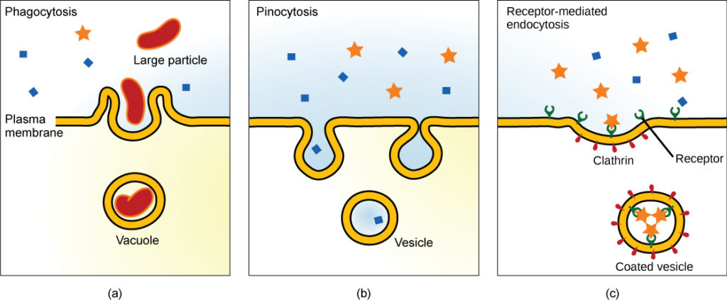
Endocytosis moves particles, such as large molecules, parts of cells, and even whole cells, into a cell. There are three types of endocytosis. Phagocytosis is the process by which large particles, such as cells, are taken in by a cell. Pinocytosis is a variation of endocytosis that means “cellular drinking”. This process takes in solutes that the cell needs from the extracellular fluid. Receptor-mediated endocytosis is a targeted variation of endocytosis that employs binding proteins in the plasma membrane that are specific for certain substances. The particles bind to the proteins and the plasma membrane invaginates, bringing the substance and the proteins into the cell. The following diagram outlines the three types of endocytosis described above. It can help you understand these forms of membrane transport by visualizing each process.
The following activity tests the reader on their understanding of the three types of endocytosis. Three real-life examples are provided for each type and the reader must drag the examples to the correct endocytic mechanism. By providing real-life examples of phagocytosis, pinocytosis, and receptor-mediated endocytosis, the reader gains a better understanding of when and where these mechanisms are used in an organism. This lays foundational knowledge that students must comprehend in order to look at larger systems.
Diffusion
Diffusion is the passive (doesn’t require energy expenditure) movement of substances across a plasma membrane. Molecules such as oxygen and carbon dioxide are transported via simple diffusion because they are small and uncharged. Lipids can also be transported by simple diffusion because of their hydrophobic nature. Diffusion is suitable for transporting substances across small distances and is driven by concentration gradients and electrical gradients.
Concentration gradients move substances from areas of higher concentration to areas of lower concentration, while electrical gradients move substances based on differences in charge. The combined gradient that affects a substance is called its electrochemical gradient, and it is especially important to muscle and nerve cells.
Tips from Past Students
The phrases “with the concentration gradient” and “against the concentration gradient” will likely come up several times during this course.
“With the concentration gradient” means that the substances are moving from the side of the cell with high concentration of that substance to the side of the cell with low concentration of that substance. This is the natural way that substances will move during diffusion.
“Against the concentration gradient” means that the substances are moving from the side of the cell with low concentration of that substance to the side of the cell with high concentration of that substance. When substances move against the concentration gradient, they require energy to get across the membrane.
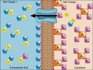
The following image provides an example of an electrochemical gradient involving sodium ions (Na+), potassium ions (K+), chloride ions (Cl–), and negatively charged proteins that are stuck inside the cytoplasm (A–). This diagram is a strong example as it clearly shows the net charge in both the extracellular fluid and the cytoplasm.
Test Your Knowledge
Thinking Beyond:
Diffusion is very important in the body for the movement of substances. For example, the movement of oxygen from the air into the blood. When would diffusion not be an appropriate tool for the body to use? Why? Hint: Think about the different properties of the molecules.
The following video provides a clear explanation of how concentration gradients and electrical gradients work. As well as how these two different types of gradients work together to form an electrochemical gradient. An electrochemical gradient can be a difficult topic for students to conceptualize. For visual-based learners, this video will enhance understanding. Chemical gradients are shown at 0:00-1:45, electrical gradients from 1:45-6:10, and electrochemical gradient and potential difference is shown at 6:10-9:40.
Test Your Knowledge
Real-Life Scenario:
There’s a grand opening of a new amusement park in your city. You’re in the crowd of people waiting to be one of the first to enter. They cut the ribbon blocking the entrance and everyone, including you, begins moving into the park. Are you moving with or against the concentration gradient? You then realize you forgot your ticket in the car and you are now trying to walk away from the park, in the opposite direction of all the people flooding in. Are you moving with or against the concentration gradient? Hint: Which is more difficult to do… possibly needing the help of energy to get you through?
Protein Channels
The cell membrane is a phospholipid bilayer, so only substances that can pass directly through the hydrophobic core can diffuse through unaided. Therefore, charged particles, which are hydrophilic by definition, and large particles use protein channels to travel across the cell membrane. Protein channels show specificity based on particle size or charge, and the number and types of channels are not fixed per cell. There are two types of protein channels.
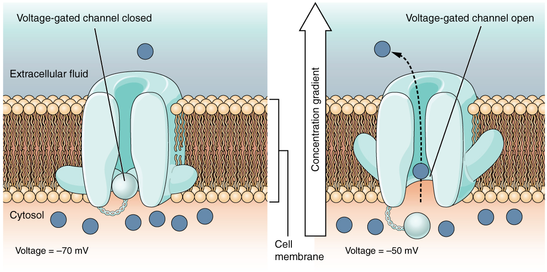
Voltage-dependent channels are a form of protein channel that responds to changes in the electrical properties of the membrane in which it is embedded. Normally, the inner portion of the membrane is at a negative voltage. When that voltage becomes less negative, the channel begins to allow ions to cross the membrane. Na+ and K+ channels in nerves and skeletal muscles are examples of voltage-dependent protein channels. The following figure illustrates the opening of a voltage-gated channel, and the movement of substances through it.
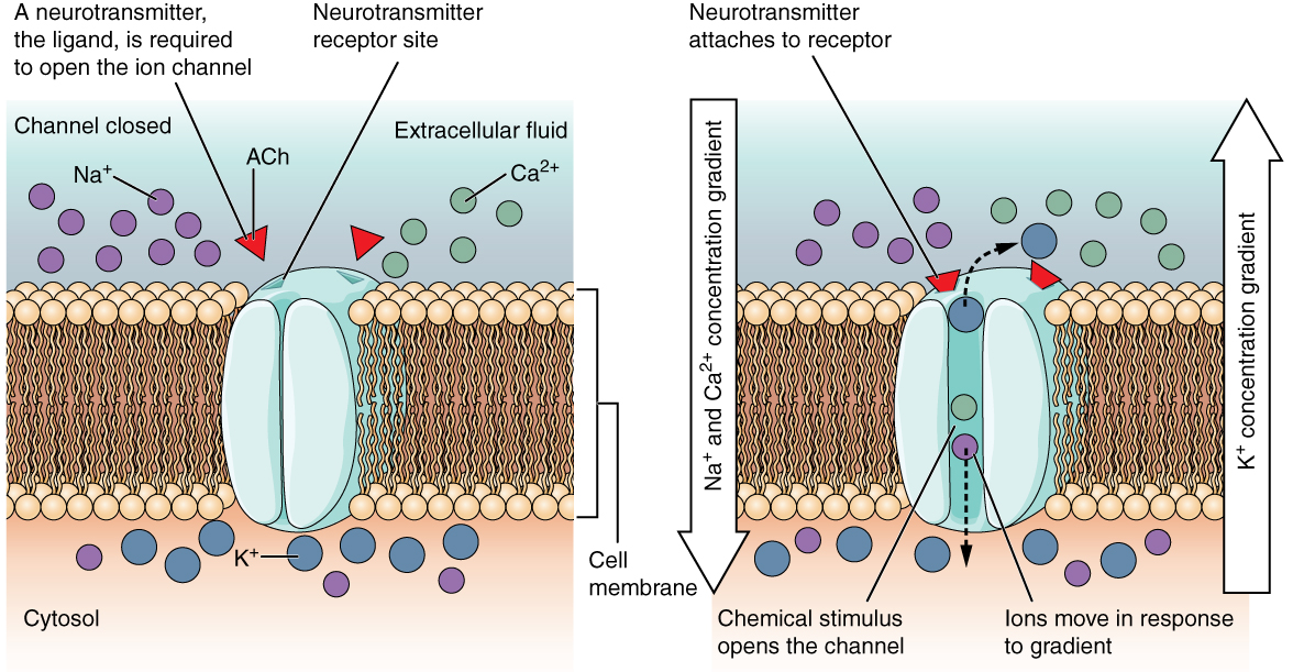
Ligand-dependent channels are a form of protein channel that opens in response to a signaling molecule, a ligand, which binds to an active site on the channel. This type of channel is also known as an ionotropic receptor because when the ligand, known as a neurotransmitter in the nervous system, binds to the protein, ions cross the membrane changing its charge. Nicotinic acetylcholine channels in the neuromuscular junction are examples of ligand-dependent protein channels. The following figure shows the mechanism behind opening a ligand channel and the movement of ions through the channel. This figure contrasts figure 3, which shows the mechanism behind voltage-gated channels.
Test Your Knowledge
Thinking Beyond:
Protein channels span across the entire cellular membrane. From what you know about hydrophilic and hydrophobic properties, which side of the protein (hydrophobic or hydrophilic) would be on the inside of the cell and which would be on the outside of the membrane? Hint: Think about where the hydrophobic and hydrophilic parts of the cell membrane are.
Active Transport
Active transport is the movement of substances from an area of low concentration to high concentration (ie. against the concentration gradient) through the use of energy such as ATP. Active transport is carried out by a specific type of transmembrane proteins called carrier proteins. There are two types of active transport.
Primary (1o) Active Transport: The movement of substances across the plasma membrane, creating a difference in charge across the membrane. This type of active transport is directly dependent on ATP.
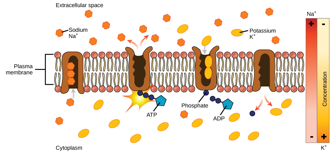
The sodium-potassium pump is a notable example of primary active transport. The pump is one of the most important in the cell, as it maintains the electrochemical gradient (and the correct concentrations of Na+ and K+). The sodium-potassium pump moves K+ into the cell while moving Na+ out at the same time, at a ratio of three Na+ for every two K+ ions moved in. The Na+-K+ ATPase exists in two forms, depending on its orientation to the interior or exterior of the cell and its affinity for either sodium or potassium ions.
The video below shows the sodium-potassium pump in action. The way that the video shows shapes within the pump that correspond to the shapes of Na+ and K+ is helpful for remembering that carrier proteins are specific for certain ions or molecules.
The diagram below shows the process of primary active transport. This example shows a sodium-potassium pump (Na+-K+ ATPase), which is one of the most important pumps in animal cells. The diagram also clearly shows that the ions are moving against their concentration gradients (from low to high concentration).
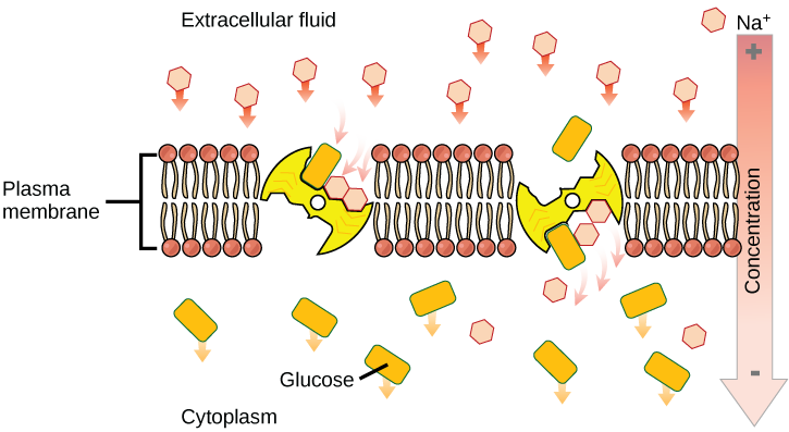
Secondary (2o) Active Transport: The movement of substances across the membrane using the electrochemical gradient established by primary active transport. This type of active transport does not directly require ATP.
The following diagram shows an example of secondary active transport using sodium ions (Na+) and glucose. The transporter moves Na+ with its concentration gradient (from high concentration to low concentration) and at the same time couples glucose movement against its concentration gradient (from low concentration to high concentration).
Still a bit confused about the difference between passive and active transport? Check out Biology: Cell Transport which provides a nice visualization of the two mechanisms.
Want some more information about secondary active transport? Check out Detailed Animation of Secondary Active Transport which helps explain how secondary active transport works by giving multiple examples.
Carrier Proteins for Active Transport
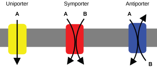
A carrier protein is a glycoprotein embedded in the plasma membrane. When a specific substance binds to a carrier protein, the protein undergoes a conformational change, resulting in the movement of the bound substance across the cell membrane. Unlike ion channels, carrier proteins use active transport. There are three types of carrier proteins. A uniporter carries one specific molecule. A pb_glossary id=”3784″]symporter[/pb_glossary] carries two different molecules in the same direction. Lastly, an pb_glossary id=”3785″]antiporter[/pb_glossary]: carries two different molecules in opposite directions.
Tips from Past Students
Membrane Transport is only a small portion of this unit. Therefore, we have decided to only include brief descriptions of each type of transport. Do not get caught up with the small details of each type – the information provided in lecture and your background information from other courses will likely be sufficient.
Did you know there is a third type of active transport? It uses photon energy (from light) to move ions and molecules across cell membranes. These light-driven pumps are commonly found in bacteria.
Test Your Knowledge
Thinking Beyond:
A central theme in HK*2810 is communication: how cells in the body ‘talk’ to each other. What do you think the cell membrane and membrane transport contribute to cell-cell communication? Hint: What might need to enter into a cell to facilitate communication?
Let’s Review! The following video provides a summary of the membrane transport section. At 0:48 ion channels for passive transport are reviewed. From 2:12 ion pumps for active transport are shown and at 2:30 secondary transporters are reviewed.
Still in need of a quick section summary? Check out this video reviewing Cell Transport!
Test Your Knowledge
Thinking Beyond:
Cell membranes are selectively permeable. This means that the membrane will only allow certain molecules or ions to pass through it. What would happen if the cell membrane suddenly lost its selective permeability? Would all substances go through it? If so, why would this be a problem? Hint: Think about homeostasis.
Key Takeaways
Consider the following concepts to help guide your studies:
- Endocytosis moves particles into a cell. The three types are pinocytosis, phagocytosis, and receptor-mediated endocytosis.
- Diffusion is the passive movement of molecules across a cell membrane.
- Concentration gradients move substances from areas of higher concentration to areas of lower concentration, while electrical gradients move substances based on differences in charge. Combined, these make up the electrochemical gradient.
- Protein channels facilitate the movement of molecules along the concentration gradient and without using any other energy source. The two types are voltage-gated channels and ligand-gated channels.
- Active transport is the movement of substances against the concentration gradient through the use of energy such as ATP. The two types are 1o active transport and 2o active transport.
- The sodium-potassium pump maintains the concentration gradient across a cell and is an example of 1o active transport
- Carrier proteins actively transport substances. The three types are uniporters, symporters, and antiporters.
Subchapter Quiz
The questions below can be used to assess your knowledge within this chapter. There are five multiple-choice questions that you should attempt without referring to your notes. The questions will provide you with responses to your answers to guide your studying but should not be used as your only resource.
Media Attributions
- Lumen Learning Endocytosis Diagram © Mariana Ruiz Villarreal is licensed under a CC BY (Attribution) license
- Electrochemical Gradients © Affordable Learning LOUIS is licensed under a CC BY-NC-SA (Attribution NonCommercial ShareAlike) license
- Voltage Gated Channels Rice University © J. Gordon Betts, Kelly A. Young, James A. Wise, Eddie Johnson, Brandon Poe, Dean H. Kruse, Oksana Korol, Jody E. Johnson, Mark Womble, Peter DeSaix is licensed under a CC BY (Attribution) license
- Ligand Gated Channels Rice University © J. Gordon Betts, Kelly A. Young, James A. Wise, Eddie Johnson, Brandon Poe, Dean H. Kruse, Oksana Korol, Jody E. Johnson, Mark Womble, Peter DeSaix is licensed under a CC BY (Attribution) license
- Primary Active Transport from CNX © Mariana Ruiz Villareal is licensed under a CC BY (Attribution) license
- Secondary Active Transport CNX © Mariana Ruiz Villareal is licensed under a CC BY (Attribution) license
- Carrier-Proteins-CNX © Lupask is licensed under a CC BY (Attribution) license
The process by which particles are moved within a cell.
The non-specific uptake of large particles into a cell.
The non-specific uptake of solutes and water from the extracellular fluid.
The uptake of specific molecules via binding to receptors on the plasma membrane and formation of vesicles intracellularly.
Net movement from high concentration to low concentration.
The tendency to repel or fail to mix with water.
The difference in the concentration of specific molecules on different sides of a cell membrane.
A difference in charge across a cell membrane.
The combination of the electrical and chemical gradients across a cell membrane.
A thin polar membrane made of two layers of lipid molecules.
A protein that allows transport of specific substances across a cell membrane.
Protein channels that are activated by changes in the electrical membrane potential near the channel.
Protein channels that allow substances to pass through in response to binding of a chemical messenger (ex. a neurotransmitter).
The movement of molecules across a cell membrane against the concentration gradient (from low concentration to high concentration), which requires the use of energy.
Adenosine 5'-triphosphate, or ATP, is the principal molecule for storing and transferring energy in cells.
A type of cell membrane protein involved in facilitated diffusion and active transport of substances across the membrane.
Movement of substances across a cell membrane using ATP for energy.
An important membrane transporter that uses primary active transport to move sodium ions and potassium ions against their concentration gradients (from areas of low concentration to high concentration).
A form of active transport across a cell membrane where a carrier protein couples the movement of a substance with its concentration gradient to the movement of another substance against its concentration gradient.
A carrier protein that transports one specific molecule across the cell membrane.
A carrier protein that transports two different molecules in the same direction across the cell membrane.
A carrier protein that transports two different molecules in opposite directions across the cell membrane.
