Regulation of Fluid Volume
The major hormones influencing total body water levels are antidiuretic hormone, aldosterone, and atrial natriuretic peptide. Circumstances that lead to fluid depletion in the body include blood loss and dehydration. Homeostasis requires that volume and osmolarity be preserved. Blood volume is important in maintaining sufficient blood pressure, and there are non-renal mechanisms involved in its preservation, including vasoconstriction, which can act within seconds of a pressure drop. Thirst mechanisms are also activated to promote the consumption of water lost through respiration, evaporation, or urination. Hormonal mechanisms are activated to recover volume while maintaining a normal osmotic environment. These mechanisms act principally on the kidney and will be discussed in this section. Keep in mind, a lot of content in this section is beyond the scope of HK*3810. Focus on the content related to the kidney and challenge yourself by considering how some of these interventions would affect the processes you have discussed in class. If you are not feeling confident with all of the material yet or are not looking for extra information, skip ahead to the next unit on hormonal regulation at the kidney.
Learning Outcomes
In this section you will learn…
- Volume sensing mechanisms of the kidney.
- The regulation of ions within the kidney.
- The elimination of drugs and hormones.
- Regulation of water balance.
- Endocrine release and regulation of blood volume.
Volume Sensing Mechanism
The body cannot directly measure blood volume, but blood pressure can be measured by specialized receptors. Blood pressure often reflects blood volume and is measured by baroreceptors in the aorta and carotid sinuses. When blood pressure increases, baroreceptors send more frequent action potentials to the central nervous system, leading to widespread vasodilation to reduce this added pressure. Included in this vasodilation, are the afferent arterioles which are connected to the glomerulus, resulting in increased glomerular filtration rate (GFR) and water loss by the kidneys. If pressure decreases, fewer action potentials travel to the central nervous system, resulting in more sympathetic stimulation resulting in vasoconstriction, which will result in decreased GFR, and water loss.
Decreased blood pressure is sensed by the granular cells in the afferent arterioles of the juxtaglomerular apparatus. In response, the enzyme renin is released. Renin activity leads to an almost immediate rise in blood pressure as renin converts angiotensinogen to activated angiotensin I, which is converted to angiotensin II. Angiotensin II causes vasoconstriction. The rise in pressure is sustained by the aldosterone effects initiated by angiotensin II; this includes an increase in sodium retention and water volume.
Cardiomyocytes of the atria also respond to a greater stretch as blood pressure rises by secreting atrial natriuretic peptide (ANP). The ANP opposes the action of aldosterone by inhibiting the recovery of sodium by the distal convoluted tubule and collecting ducts. More sodium is lost, and as water follows, total blood volume and pressure decline. In low-pressure states, ANP does not have much of an effect. The video below is useful if you need clarification or more information regarding the hormones that act within the body to regulate blood pressure. [1]
Test Your Knowledge
Clinical Application:
The most frequently prescribed anti-hypertensive diuretic is hydrochlorothiazide. It inhibits the Na+/ Cl– symporter in the DCT and collecting duct. How might this affect water movement in the nephron? Hint: Where sodium goes, water flows!
Regulation of Ions
Regulation of Extracellular Sodium
Sodium has a very strong osmotic effect and attracts water. It plays a larger role in the osmolarity of the plasma than any other circulating component of the blood. If there is too much sodium present, either due to poor control or excess dietary consumption, a series of metabolic problems occur. There is an increase in the total volume of water that travels into the bloodstream to dilute the increased sodium levels, which leads to hypertension. Over a long period, this increases the risk of serious complications such as heart attacks, strokes, and aneurysms. It can also contribute to system-wide edema.
Mechanisms for regulating sodium concentration include the renin-angiotensin-aldosterone system and antidiuretic hormone (ADH). Aldosterone stimulates the uptake of sodium on the apical cell membrane of cells in the distal collecting tubule and collecting ducts, whereas ADH helps to regulate sodium concentration indirectly by regulating the reabsorption of water.
Regulation of Extracellular Potassium
Potassium is present in a 30-fold greater concentration inside the cell than outside the cell. A generalization can be made that potassium and sodium concentrations will move in opposite directions due to their respective concentration gradients. When more sodium is reabsorbed, more potassium is secreted; when less sodium is reabsorbed leading to excretion by the kidney, more potassium is retained. When aldosterone causes a recovery of sodium in the nephron, a negative electrical gradient is created which promotes the secretion of potassium and chloride into the lumen.
Regulation of Chloride
Chloride is important in acid–base balance in the extracellular space and has other functions, such as in the stomach, where it combines with hydrogen ions in the stomach lumen to form hydrochloric acid, aiding digestion. Its close association with sodium in the extracellular environment makes it the dominant anion of this compartment, and its regulation closely mirrors that of sodium.
Regulation of Calcium and Phosphate
The parathyroid glands monitor and respond to circulating levels of calcium in the blood. When levels drop too low, parathyroid hormone (PTH) is released to stimulate the distal convoluted tubule to reabsorb calcium from the forming urine. When levels are adequate or high, less PTH is released and more calcium remains in the forming urine to be lost. Phosphate levels move in the opposite direction. When calcium levels are low, PTH inhibits the reabsorption of phosphate so that its blood level drops, allowing calcium levels to rise. PTH also stimulates the renal conversion of calcidiol into calcitriol, the active form of vitamin D. Calcitriol then stimulates the intestines to absorb more calcium from the diet.
Regulation of Nitrogen Waste
Nitrogen wastes are produced by the breakdown of proteins during normal metabolism. Proteins are broken down into amino acids, which in turn are deaminated by having their nitrogen groups removed. Deamination converts the amino (NH2) groups into ammonia (NH3), ammonium ion (NH4+), urea, or uric acid. Ammonia is extremely toxic, so most of it is very rapidly converted into urea in the liver. Human urinary wastes typically contain primarily urea with small amounts of ammonium and very little uric acid.
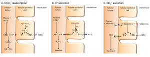
Regulation of Hydrogen Ion, Bicarbonate, and pH
H2O + CO2 ⇔ H2CO3 ⇔ H+ + HCO3–
The acid–base homeostasis of the body is a function of chemical buffers provided by the lungs and kidneys. Buffers, especially proteins, bicarbonate, and ammonia have a very large capacity to absorb or release hydrogen ions as needed to resist a change in pH. They can act within fractions of a second. The lungs can rid the body of excess acid very rapidly within seconds to minutes through the conversion of bicarbonate into carbon dioxide, which is then exhaled. It is rapid but has a limited capacity in the face of a significant acid challenge. The kidneys can rid the body of both acid and base. The renal capacity is large but slow-acting within minutes to hours. The cells of the proximal convoluted tubule actively secrete hydrogen into the forming urine as sodium is reabsorbed. The body rids itself of excess hydrogen ions and raises blood pH. In the collecting ducts, the apical surfaces of intercalated cells have proton pumps that actively secrete hydrogen into the luminal, forming urine to remove it from the body. As hydrogen ions are pumped into the forming urine, it is buffered by bicarbonate, dihydrogen phosphate ion, or ammonia (forming NH4+, ammonium ion). Urine pH typically varies in a normal range from 4.5 to 8.0. The concept of ionic regulation in the kidneys is shown below in figure 1. [2]
Tips From Past Students
Remember, these processes can occur all at the same time in various parts of the kidney!
Elimination of Drugs and Hormones
Water-soluble drugs may be excreted in the urine and are influenced by one or all of the following processes: glomerular filtration, tubular secretion, or tubular reabsorption. Structurally small drugs can be filtered by the glomerulus with the filtrate. Large drug molecules such as heparin or those that are bound to plasma proteins cannot be filtered and are not readily eliminated. Some drugs can be eliminated by carrier proteins that enable the secretion of the drug into the tubule lumen. There are specific carriers that eliminate basic drugs such as dopamine or histamine and acidic drugs such as penicillin or indomethacin. As is the case with other substances, drugs may be both filtered and reabsorbed passively along a concentration gradient.[3]
Water Balance
On a typical day, the average adult will consume around 2.5 L of aqueous fluids. Although most of the intake comes through the digestive tract, about 230 mL per day is generated metabolically, in the last steps of aerobic respiration. Additionally, each day about the same volume of water leaves the body by different routes; most of which is excreted in the urine. The kidneys also can adjust blood volume through mechanisms that draw water out of the filtrate and urine. Due to this, the kidneys can contribute to the regulation of water levels in the body; they conserve water if you are dehydrated, and they can make the urine more dilute to expel excess water if necessary. Water can also be lost through the skin utilizing evaporation from the skin surface and air expelled from the lungs. This type of water loss is called insensible water loss because a person is usually unaware of it.[4]
Test Your Knowledge
Thinking Beyond:
If adequate fluids are not consumed, dehydration results and a person’s body contains too little water to function correctly. A person who repeatedly vomits or who has diarrhea may become dehydrated, and infants, because their body mass is so low, can become dangerously dehydrated very quickly. Endurance athletes such as distance runners often become dehydrated during long races. Dehydration can be a medical emergency, and a dehydrated person may lose consciousness, become comatose, or die if his or her body is not rehydrated quickly. What renal compensations would occur if you were dehydrated? Hint: Consider the effects that blood volume and pressure would have on hormonal release.
Regulation of Water Intake
Plasma Osmolality
Plasma osmolality represents the ratio of solutes to water in blood plasma. A person’s plasma osmolality value reflects his or her state of hydration. A healthy body maintains plasma osmolality within a narrow range, by employing several mechanisms that regulate both water intake and output. Drinking water is considered voluntary. So how is water intake regulated by the body? Consider someone who is experiencing dehydration, a net loss of water that results in insufficient water in the blood and other tissues. The water that leaves the body, as exhaled air, sweat, or urine, is ultimately extracted from blood plasma. As the blood becomes more concentrated, the thirst response—a sequence of physiological processes—is triggered. The resource below summarizes the overall balance of water in the body and introduces the concept of thirst response.
Thirst Response
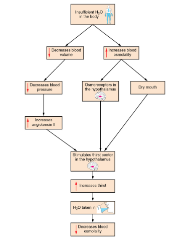
Osmoreceptors, sensory receptors in the thirst centre of the hypothalamus, monitor the concentration of solutes in the blood. If blood osmolality increases above its ideal value, the hypothalamus transmits signals that result in conscious awareness of thirst. The person should, and normally does, respond by drinking water. The hypothalamus of a dehydrated person also releases antidiuretic hormone (ADH) through the posterior pituitary gland. ADH signals the kidneys to recover water from urine, effectively diluting the blood plasma. To conserve water, the hypothalamus also signals via the sympathetic nervous system to the salivary glands in the mouth. The signals result in a decrease in watery, serous output and an increase in stickier, thicker mucus output. These changes in secretions result in the feelings of “dry mouth” and the sensation of thirst. The thirst response is depicted in the flowchart seen below in figure 2, this learning object is beyond the HK*3810 course level.[5]
Additional effects of decreased blood volume :
1. Increase Heart Rate and Strength
Baroreceptors found in the arch of the aorta and the carotid arteries in the neck, detect a decrease in blood pressure that results from decreased blood volume. The heart is ultimately signalled to increase its rate and/or strength of contractions to compensate for the lowered blood pressure.
2. Stimulate renin-angiotensin system
Kidneys have a renin-angiotensin hormonal system that increases the production of the active form of the hormone angiotensin II, which helps stimulate thirst but also stimulates the release of the hormone aldosterone from the adrenal glands. Aldosterone increases the reabsorption of sodium in the distal tubules of the nephrons in the kidneys, and water follows this reabsorbed sodium back into the blood.[6]
Regulation of Water Output
The kidneys must also make adjustments in the event of the ingestion of too much fluid. Diuresis, which is the production of urine in excess, begins about 30 minutes after drinking a large quantity of fluid. Diuresis reaches a peak after about one hour, and normal urine production is reestablished after about three hours. Did you know that water loss from the body occurs predominantly through the renal system? A person produces an average of 1.5 litres of urine per day. Although the volume of urine varies in response to hydration levels, there is a minimum volume of urine production required for proper bodily functions. The kidney excretes 100 to 1200 milliosmoles of solutes per day to rid the body of a variety of excess salts and other water-soluble chemical wastes, most notably creatinine, urea, and uric acid. Failure to produce the minimum volume of urine means that metabolic wastes cannot be effectively removed from the body, a situation that can impair organ function. The minimum level of urine production necessary to maintain normal function is about 0.47 litres per day. [7]
Key Takeaways
Consider the following concepts to help guide your studies:
- How the kidney responds to a drop in blood pressure.
- The location and hormone dependence of ion regulation.
- How the kidney regulates fluid flux.
Sub-chapter Quiz
The questions below can be used to assess your knowledge within this chapter. There are five multiple-choice questions that you should attempt without referring to your notes. The questions will provide you with responses to your answers to guide your studying but should not be used as your only resource.
Media Attributions
- Bicarbonate Regulation Kidney © Natalie's Casebook: FOAM is licensed under a CC BY (Attribution) license
- Dehydration Integration © BC Campus is licensed under a CC BY (Attribution) license
- 25.9 Regulation of Fluid Volume and Composition - Anatomy and Physiology. OpenStax. (2013, April 25). ↵
- 25.9 Regulation of Fluid Volume and Composition - Anatomy and Physiology. OpenStax. (2013, April 25). ↵
- 25.9 Regulation of Fluid Volume and Composition - Anatomy and Physiology. OpenStax. (2013, April 25). ↵
- 26.2 Water Balance - Anatomy and Physiology. OpenStax. (2013, April 25). ↵
- 26.2 Water Balance - Anatomy and Physiology. OpenStax. (2013, April 25). ↵
- 26.2 Water Balance - Anatomy and Physiology. OpenStax. (2013, April 25). ↵
- 26.2 Water Balance - Anatomy and Physiology. OpenStax. (2013, April 25). ↵
A hormone that is released from the posterior pituitary gland in response to low blood pressure. ADH increases water reabsorption in the kidney, which increases blood volume, and decreases urine excretion.
A hormone that is released from the adrenal cortex in response to angiotensin II or in direct response to an increased plasma K+ concentration. It promotes Na+ reabsorption by the nephron, promoting the retention of water.
A peptide hormone released from the heart that promotes the excretion of water and sodium by reducing the function of ALDO, ADH, and renin.
Sensory receptor cell that is sensitive to changes in pressure (detected through stretch).
An area within the carotid artery that contains important baroreceptors.
Relaxation of smooth muscle cells and the widening of blood vessels. This will allow for more blood flow through the vasculature.
The volume of filtrate formed by both kidneys per minute.
Contraction of smooth muscle cells and the tightening of blood vessels. This will allow for less blood flow through the vasculature.
A structure formed by the distal convoluted tubule and the glomerular afferent arterioles. Involved in the regulation of blood pressure and GFR.
An enzyme produced by the granular cells of the afferent arteriole at the JGA. It enzymatically converts angiotensinogen to angiotensin I.
Cells within the heart responsible for generating force and allowing the heart to contract rhythmically.
Any substance that promotes diuresis (the increased production of urine), which also promotes water excretion.
A hormone secreted by the parathyroid glands that plays a role in regulating blood calcium concentration.
The process of removing a substance out of the bloodstream and into the urine.
In terms of kidney physiology, reabsorption is when the kidney removes water and solutes from the nephron and returns them to circulation.
A drug used to prevent the formation of blood clots.
The cellular process of producing energy (ATP) including glycolysis, the citric acid cycle, and the electron transport system.
Small portion of the brain that regulates metabolic processes, serving as a link between endocrine and nervous systems via the pituitary gland.
Prepares the body to respond to stress, such as a threat or injury. It is often referred to as the “fight-or-flight” response.
A potent vasoconstrictor that plays an immediate role in the regulation of blood pressure.
The increased production of urine.
